How a VQ Scan Helps Diagnose Lung Conditions
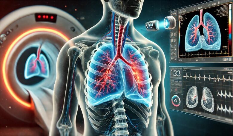
In the field of pulmonary diagnostics, the VQ scan stands out as a crucial imaging tool for diagnosing various lung conditions. The VQ scan, or ventilation-perfusion scan, offers detailed insights into the airflow and blood flow within the lungs, helping healthcare professionals identify issues such as pulmonary embolism and other respiratory disorders. This article explores the importance of the VQ scan, its applications, and its role in enhancing lung health diagnosis.
Understanding the VQ Scan
A VQ scan is a type of nuclear medicine test that evaluates ventilation (airflow) and perfusion (blood flow) in the lungs. It comprises two parts:
Ventilation Scan: This part of the test assesses how air moves through the lungs. The patient inhales a slightly radioactive gas or aerosol, and a special camera captures images of the airflow patterns.
Perfusion Scan: This segment evaluates blood flow in the lungs. A radioactive tracer is injected into a vein, and images are taken to show how well blood circulates throughout the lung tissue.
By comparing the ventilation and perfusion images, doctors can identify discrepancies that might indicate issues such as blockages or abnormalities in lung function.
Applications of the VQ Scan
Diagnosing Pulmonary Embolism
One of the primary uses of a VQ scan is diagnosing pulmonary embolism (PE), a serious condition where a blood clot blocks blood flow in the lungs. The VQ scan is particularly useful when other imaging techniques, such as a CT pulmonary angiogram, are not suitable due to allergies or kidney issues.
Detecting Blood Clots: By highlighting areas with normal ventilation but reduced perfusion, the VQ scan can pinpoint the location of blood clots obstructing blood flow.
Non-Invasive and Safe: The VQ scan is a non-invasive procedure, making it a safer option for patients unable to undergo more invasive tests.
Evaluating Lung Function in Chronic Conditions
VQ scans are also used to assess lung function in patients with chronic respiratory conditions such as chronic obstructive pulmonary disease (COPD) or emphysema.
Monitoring Disease Progression: The scans provide valuable information about changes in lung function over time, aiding in the management of chronic lung diseases.
Assessing Surgical Risks: In patients undergoing lung surgery, VQ scans help evaluate the risks and ensure that the remaining lung tissue can adequately support breathing postoperative.
Post-Surgical and Transplant Evaluation
After lung surgery or transplantation, a VQ scan can be employed to monitor the success of the procedure and the functionality of the transplanted or remaining lung tissue.
The VQ Scan Procedure
The VQ scan is a simple, outpatient procedure that typically takes about 30 to 60 minutes. Here’s what patients can expect:
Preparation: No special preparation is usually required, though patients may be asked to remove jewelry or metal objects. It’s important to inform the healthcare provider of any medications being taken or allergies to contrast materials.
Ventilation Scan: The patient inhales a radioactive gas through a mask or mouthpiece, and images of the lungs are taken using a gamma camera.
Perfusion Scan: A small amount of radioactive tracer is injected into a vein, and further images are captured to show blood flow patterns in the lungs.
Analysis: The images from the ventilation and perfusion scans are compared to identify any ventilation-perfusion mismatches, which can indicate lung problems.
Safety and Considerations
The VQ scan is generally safe and involves minimal exposure to radiation, similar to that of a standard chest X-ray. However, it is not recommended for pregnant women due to the potential risk to the fetus. Patients should discuss all health conditions and concerns with their doctor prior to the test.
Advancements and Future Prospects
Advancements in VQ scan technology continue to improve its accuracy and reliability. Newer imaging techniques and software developments are enhancing the ability to interpret scan results more precisely, making the VQ scan an even more valuable tool in pulmonary diagnostics.
Additionally, research is ongoing to expand the applications of VQ scans in diagnosing other lung conditions and to integrate them with other imaging modalities for comprehensive lung assessments.
Conclusion
The VQ scan is an essential diagnostic tool for evaluating lung conditions, providing critical insights into both ventilation and perfusion within the lungs. Its ability to non-invasively detect conditions like pulmonary embolism, assess lung function, and monitor post-surgical outcomes makes it invaluable in modern respiratory care. As technology advances, the role of VQ scans in diagnosing and managing lung health is poised to grow, offering patients a path to earlier detection and more effective treatment of lung conditions.