Proximal Humeral Nail Systems: Technique, Stability, and Clinical Outcomes
Fractures near the top of the arm bone, just below the shoulder joint, are surprisingly frequent. They happen in elderly patients with thinning bones after a simple fall at home, and in younger people after road traffic accidents or sports injuries. The tricky part is not just the break itself but the fact that the upper humerus is wrapped in crucial muscles and relies on a delicate blood supply. Getting this region to heal properly is no small task for surgeons.
Over the years, different fixation methods have been tried—plates, screws, even arthroplasty in severe cases. But one solution that has steadily grown in popularity is the proximal humeral nail system. Surgeons favor it because it combines strength with a relatively gentle surgical approach, sparing much of the soft tissue that other methods disturb.
The Surgical Routine in Practice
In the operating theatre, the setup looks fairly standard. Patients are placed in what is called the beach-chair position—basically half-sitting, half-lying—which allows the surgeon good visibility. Imaging is critical, so a C-arm fluoroscope hovers nearby to guide every step.
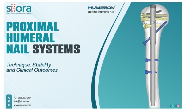
Access is gained through a small incision at the top of the shoulder. This is not a “big exposure” operation; the idea is to disturb as little as possible. The guidewire is carefully passed into the bone canal, and reaming creates just enough room for the nail. Every move is checked on live X-ray images. Once the nail is inserted, screws are locked into the bone fragments around the humeral head. These give rotational stability and prevent collapse. A couple of screws are added lower down the shaft to secure the construct. All of this typically takes less time than applying a plate, and the incision is much smaller.
Why Nails Provide the Needed Stability?
The science behind the nail’s stability is fairly straightforward. Because it sits inside the bone, right along its central load line, it shares stress in a way that feels very “natural” for the humerus. Plates, on the other hand, sit on the outside, which means they often need a greater area of bone exposure. The intramedullary position coupled with multiple locking options gives the nail a biomechanical edge, especially in osteoporotic bone.
Modern designs also address older complications. Multiple screw angles help catch firmer parts of the head, reducing risks of screws cutting out. This has been particularly helpful for elderly women with weaker bone quality, where conventional fixation tends to fail more often.
What Studies and Clinical Use Show?
Most published data point to high union rates—well above 90 percent in many series. Healing is usually achieved within a few months, though age and fracture complexity certainly influence recovery time. Functionally, patients tend to regain a decent arc of movement, and pain levels are reliably reduced compared to pre-surgery status.
That said, outcomes are not flawless. Surgeons still encounter problems like impingement from misplaced screws or irritation around the nail entry point. With experience and improved orthopedic implants, however, these complications are becoming less frequent.
When placed side by side with locking plates, nails demonstrate several advantages: smaller incisions, shorter surgical time, and less blood loss. Plates, admittedly, can be superior when the fracture is extensively comminuted and anatomic reconstruction is essential. Thus, the choice isn’t always straightforward; it depends heavily on the individual fracture and the surgeon’s judgment.
A Balanced Perspective
What makes proximal humeral nails appealing is not that they are perfect but that they strike a balance—strong internal fixation without the excessive disruption that older techniques often required. For an elderly patient, that means fewer surgical stresses, lower infection risks, and more predictable recovery. For the surgeon, it means a reliable technique backed by good long-term data.